 Contact Us Today
 Begin My Assessment
 Watch Us On Youtube
 enquiries@spineconnection.org


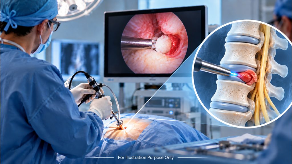
A small piece of disc can press on a nerve and can be life changing. This pressure often causes sciatica, sharp leg pain, numbness, tingling, or weakness that makes walking and sitting hard. Endoscopic microdiscectomy targets this exact problem. Surgeons remove only the small disc fragment pressing on your nerve, leaving the rest of your healthy disc alone.
The procedure uses one tiny cut, a thin camera called an endoscope, and small tools built for precise, careful work. Instead of a large opening and a long recovery, this approach focuses on one goal. Free your nerve, ease your pain, and get you back on your feet quickly.
What Is Endoscopic Microdiscectomy?
Endoscopic microdecompression, also called full-endoscopic lumbar discectomy, is a minimally invasive procedure for a herniated disc pressing on a spinal nerve.
Your surgeon makes a small opening in your skin and guides a thin endoscope toward the affected disc. A high-definition camera at the end of the scope sends a clear, magnified view to a screen. Your surgeon uses this view to see the nerve and the herniated disc fragment.
Small surgical tools pass through the same narrow working channel. Your surgeon then removes the loose disc material pressing on the nerve.
The procedure focuses on the problem area. When suitable, your surgeon leaves the healthy part of the disc in place and avoids unnecessary disturbance of nearby muscles and soft tissue.
Why Does a Herniated Disc Cause Nerve Pain?
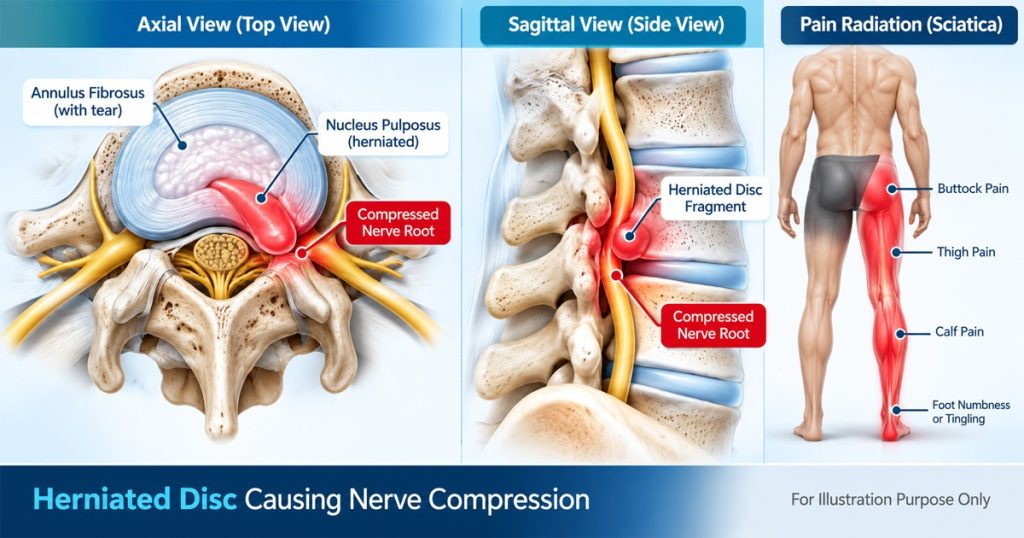
Your spinal discs sit between the bones of your spine. Each disc has a soft inner nucleus and a stronger outer annulus. When the outer layer develops a tear or weak spot, some of the soft inner tissue can move outward.
If this disc material presses on a nearby nerve, you may feel pain along the path of the nerve. With a lower back disc problem, pain often travels into the buttock and leg.
You may also experience:
- Leg pain
- Numbness
- Tingling
- Muscle weakness
This is where sciatica and pinched nerve pain often come into the picture. The symptoms result from irritation or pressure around a spinal nerve.
Endoscopic disc surgery targets the disc fragment causing this pressure. Removing the fragment gives the affected nerve more room.
How Does Endoscopic Disc Surgery Remove the Disc Fragment?
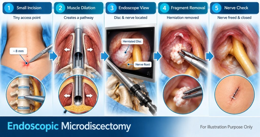
Your endoscopic spine surgery follows a careful series of steps. Here is how the whole surgery will be done.
1. A small incision
The surgeon makes a tiny skin opening, usually about 7 millimeters to 1 centimeter. This opening is smaller than a pencil eraser.
2. A narrow path reaches the disc
Special dilators gently move the muscle fibers apart. This creates a path for the endoscope and surgical tools without making a large opening through the back muscles.
3. The camera shows the treatment area
The endoscope provides a bright, magnified view of your nerve and disc. The image appears on a high-definition monitor. Fluid also helps keep the area clear during surgery.
4. The surgeon removes the loose fragment
Small instruments pass through the working channel. Your surgeon uses these tools to remove the disc fragment pressing on your nerve.
The focus stays on the problem fragment. The surgeon leaves the healthy part of your disc in place when appropriate.
5. The nerve is checked, and the incision is closed
After removing the fragment, your surgeon checks the nerve to make sure the pressure has been relieved. The instruments then come out, and the small incision is usually closed with one stitch or surgical tape.
Choosing the Access Route: Interlaminar or Transforaminal
The right route depends on where your disc fragment sits. Your surgeon reviews your MRI and other spine images before choosing the access point.
Transforaminal endoscopic approach
The Transforaminal endoscopic discectomy reaches the disc through the neural foramen. This is the small opening where a spinal nerve leaves the spine.
Interlaminar endoscopic approach
The interlaminar approach reaches the spine between two lamina bones at the back of the spine. Your surgeon may choose this route when the disc fragment sits closer to the center of the spinal canal.
The route depends on your anatomy and the location of the herniated disc. Your surgeon uses your imaging and symptoms to select the approach.
What To Choose: Microdiscectomy Or Endoscopic Discectomy
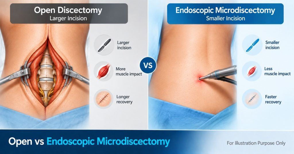
People often search for microdiscectomy vs endoscopic discectomy when they start learning about treatment options.
Both procedures remove disc material to take pressure off a spinal nerve. The main difference involves how the surgeon reaches and views the affected area.
Traditional microdiscectomy uses a small incision and a microscope. Endoscopic discectomy uses a thin endoscope with a camera. The camera sends the surgical view to a monitor.
Your symptoms, MRI findings, disc location, and overall spine condition help your surgeon decide which approach fits your case.
What Are the Potential Benefits?
Minimally invasive herniated disc surgery uses a small access point and targets the specific disc fragment causing nerve pressure.
For suitable patients, potential benefits include:
- A small incision
- Less disturbance of back muscles and nearby soft tissue
- Minimal blood loss
- A small external scar
- Less post-operative pain in many cases
- An earlier return to daily activities compared with larger open surgery
- Walking on the same day for many patients
- Going home within a few hours for many outpatient cases
Your recovery depends on your health, the location of your disc problem, the procedure, and your surgeon’s instructions. Your care team should explain what you can expect before surgery.
Who Might Be Considered for Endoscopic Disc Surgery?
Doctors often consider endoscopic disc surgery for people with symptoms linked to a herniated disc pressing on a spinal nerve.
These symptoms may include:
- Leg pain
- Numbness
- Tingling
- Weakness
Your doctor first reviews your symptoms and spine images. Surgery may enter the discussion when non-surgical treatment has not provided enough relief or when ongoing nerve pressure raises concern about nerve damage.
A herniated disc does not automatically mean you need surgery. Your surgeon looks at the full picture before recommending a procedure.
What Are the Limits and Possible Risks?
Endoscopic disc surgery focuses on removing the disc fragment pressing on the nerve. The procedure does not treat every cause of back pain.
Possible complications include recurrent disc herniation, a remaining disc fragment, a tear in the protective covering around the nerves, nerve irritation or injury, infection, and bleeding.
Some people also continue to have lower back pain after surgery. This may happen when the disc has other damage beyond the fragment pressing on the nerve.
Large, hard, or calcified disc material may require another surgical approach. Spinal instability or a structural problem may also call for a different treatment.
Your surgeon should explain the possible benefits, risks, and other treatment choices for your specific condition.
What Is Recovery Like After Endoscopic Disc Surgery?
Your endoscopic herniated disc surgery recovery often starts soon after the procedure. Many patients walk on the same day and return home within a few hours when the procedure is performed as outpatient care.
Light activity often starts within one to two weeks, based on your surgeon’s instructions. Your activity should increase step by step as your body heals.
Post-operative rehabilitation may also form part of your recovery plan. Your care team may guide you on safe movement, activity limits, exercises, and your return to normal daily tasks.
Your recovery timeline depends on your condition and how your body responds after surgery. Follow your surgeon’s instructions closely throughout the healing process.
A Focused Approach to a Herniated Disc
When a herniated disc presses on a nerve, finding the right treatment starts with understanding the exact problem. Endoscopic microdiscectomy offers a focused way to remove a loose disc fragment through a small opening.
If your MRI shows a herniated disc and your symptoms continue, Spine Connection helps you connect with experienced endoscopic spine surgeons in Germany and Thailand. We also help coordinate specialist opinions, treatment planning, hospital arrangements, travel, accommodation, and follow-up care.
If a herniated disc is affecting your daily life, start with a clear review of your condition. Begin My Assessment, or reach out through Ask Our Doctors, and take the pressure off your nerve now.
FAQs
What is endoscopic microdiscectomy?
Endoscopic microdiscectomy is a minimally invasive procedure used to remove a herniated disc fragment pressing on a spinal nerve. Your surgeon uses a small incision, a thin camera, and small surgical tools.
Is endoscopic discectomy the same as keyhole spine surgery?
Keyhole spine surgery is a common term for an endoscopic approach. The procedure uses a small opening and an endoscope to help the surgeon reach and treat the affected area.
What is the difference between transforaminal and interlaminar surgery?
A transforaminal approach reaches the disc through the opening where the spinal nerve leaves the spine. An interlaminar approach reaches the area between two lamina bones at the back of the spine. Your MRI and the location of your disc fragment help guide the choice.
How long does recovery take after endoscopic microdiscectomy?
Many patients walk on the same day. Light activity often resumes within one to two weeks, based on the surgeon’s advice. Full recovery varies from person to person and depends on the condition treated and your healing progress.

