Thank You!

Your message has been submitted successfully. We will get back to you shortly.

Cover Top Placeholder

Spinal Stenosis

Cover Middle Accent
Cover Bottom Height Engine
Last updated:

What is spinal stenosis?

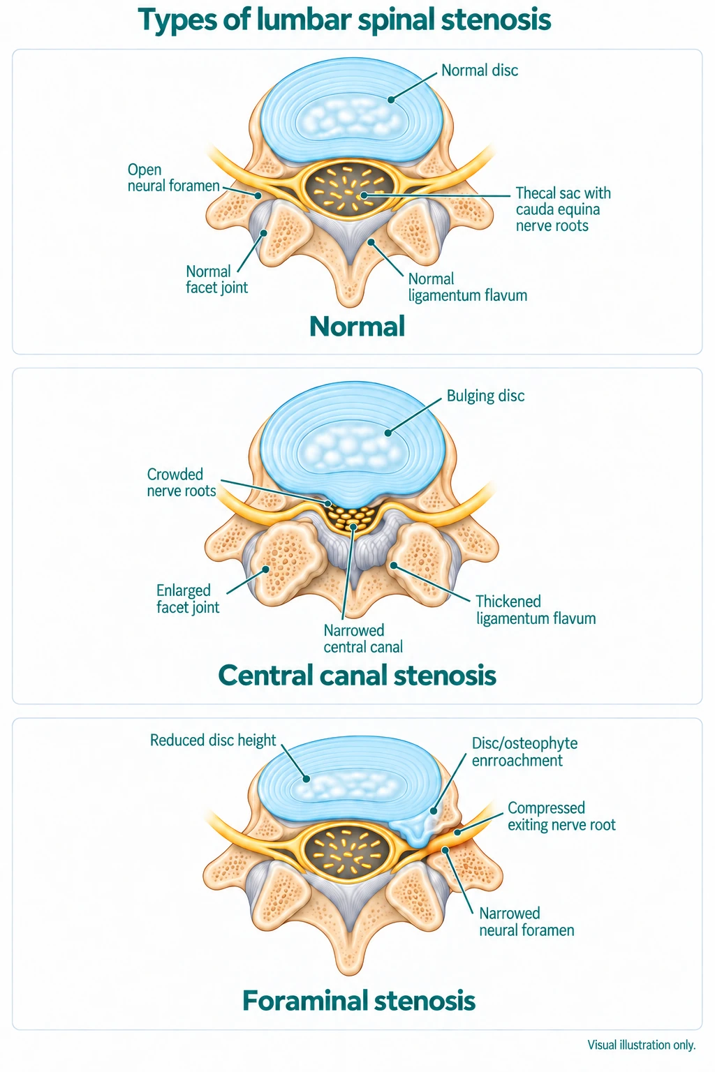
Spinal stenosis is a narrowing of the spaces within the spine, leaving less room for the spinal cord or nerve roots. This can put pressure on these structures and cause pain, numbness, tingling, or weakness. In the lower back, lumbar stenosis is one of the most common reasons older adults have spine surgery. The narrowing can be in the central canal, in the lateral recesses just off to the side, or in the openings where the nerves leave the spine (the foramina).

It is usually a gradual, age-related process rather than a sudden event, and, like other wear-related changes, it shows up on the scans of many people who have no symptoms at all. Whether it causes any symptoms, and to what degree, varies a great deal from person to person.
Patient Education Video

What does spinal stenosis feel like?

The hallmark of lumbar stenosis is neurogenic claudication: pain, heaviness, cramping, or numbness in the buttocks and legs that comes on with standing and walking, and eases when you sit down or bend forward. Many people find they can walk further leaning on a shopping trolley, or that riding a bike is comfortable while walking upright is not, because bending forward opens up the space around the nerves. Walking distance is how most people end up measuring the condition for themselves, and it varies enormously: some walk for miles with occasional heaviness, others find symptoms come on within minutes. A walking distance that is steadily shrinking is telling, and symptoms are often felt in both legs, while lower back pain may not always be present.

Some symptoms need urgent medical attention rather than watchful waiting: loss of bladder or bowel control, numbness around the groin or inner thighs, or leg weakness that is quickly getting worse. These symptoms may signal cauda equina syndrome, a medical emergency that requires urgent assessment.

What causes spinal stenosis?

The usual cause is age-related wear that adds up over time, narrowing the space around the nerves in several ways at once:

  • Disc changes. As a disc dries out and loses height, it can bulge backwards into the canal, and the loss of height also brings the vertebrae closer together. That narrows the foramina, the side openings where the nerve roots leave the spine, and can pinch a root as it passes through (foraminal stenosis). A disc that herniates can press directly into the canal or the foramen and compress a nerve on its own.
  • Facet joints. As the facet joints become arthritic they enlarge, crowding the space at the back and the sides of the canal.
  • Ligaments. The ligamentum flavum, a ligament inside the canal, thickens with age and can buckle inward, taking up more room.

Each of these takes up space, and often several act together.

Because of this, stenosis frequently travels with the other wear-related conditions: degenerative disc disease, facet arthropathy, and spondylolisthesis, where one vertebra slips forward on the one below and narrows the canal. Some people are simply born with a narrower canal than average (congenital stenosis), which means less age-related change is needed before symptoms appear.

How is spinal stenosis diagnosed?

Diagnosis starts with your history and a physical examination. The walking pattern is often the biggest clue: symptoms that build with standing and walking and settle when you sit or lean forward point strongly to stenosis. An MRI (or a CT scan) then confirms where the narrowing is and how tight it is. As with other spine conditions, the images have to be read alongside the symptoms, because narrowing is common on the scans of older people who feel perfectly well.

How severe is the narrowing?

On an MRI, spinal stenosis is often described as mild, moderate, or severe, based on how much the canal or the openings for the nerves have narrowed.

Mild: some narrowing, often with little or no effect on the nerves.

Moderate: more definite narrowing, usually with symptoms such as leg discomfort or heaviness on walking.

Severe: marked narrowing that clearly crowds the nerves, often with more constant or limiting symptoms.

As with the diagnosis, the grade on the scan and the trouble it causes do not always match. Significant narrowing can cause few symptoms, and moderate narrowing can be quite limiting. The recommended medical treatment takes into account your clinical symptoms and how you feel, not what the spine imaging shows alone.

How is spinal stenosis treated?

Most people with spinal stenosis are managed without surgery, at least to begin with. Symptoms often stay stable or fluctuate rather than marching steadily worse, so there is usually time to try conservative care first.

1. Conservative care (first line). Clinical guidelines recommend starting here: staying active, physiotherapy (including forward-bending exercises and walking programmes), weight management, and pain relief where appropriate. The movements that tend to aggravate lumbar stenosis are the ones that arch the back backwards, along with long periods standing still, while forward-leaning positions usually relieve it, which is why cycling and an inclined treadmill are often better tolerated than walking upright. No single medication stands out as the best for stenosis, and it works better as a support to activity and physiotherapy than as a treatment in its own right; what suits you is a conversation with your own doctor. Epidural steroid injections can give some people a period of relief, though the benefit is often partial and temporary.

2. Surgery. Surgery is considered when leg symptoms and walking limitation persist despite conservative care, or when there is significant or worsening weakness. The aim is to reopen the space around the nerves.

What are the surgery options for spinal stenosis?

When surgery is needed, the choice depends on how much room the nerves need and whether the segment is stable. The main options are:

1. Decompression

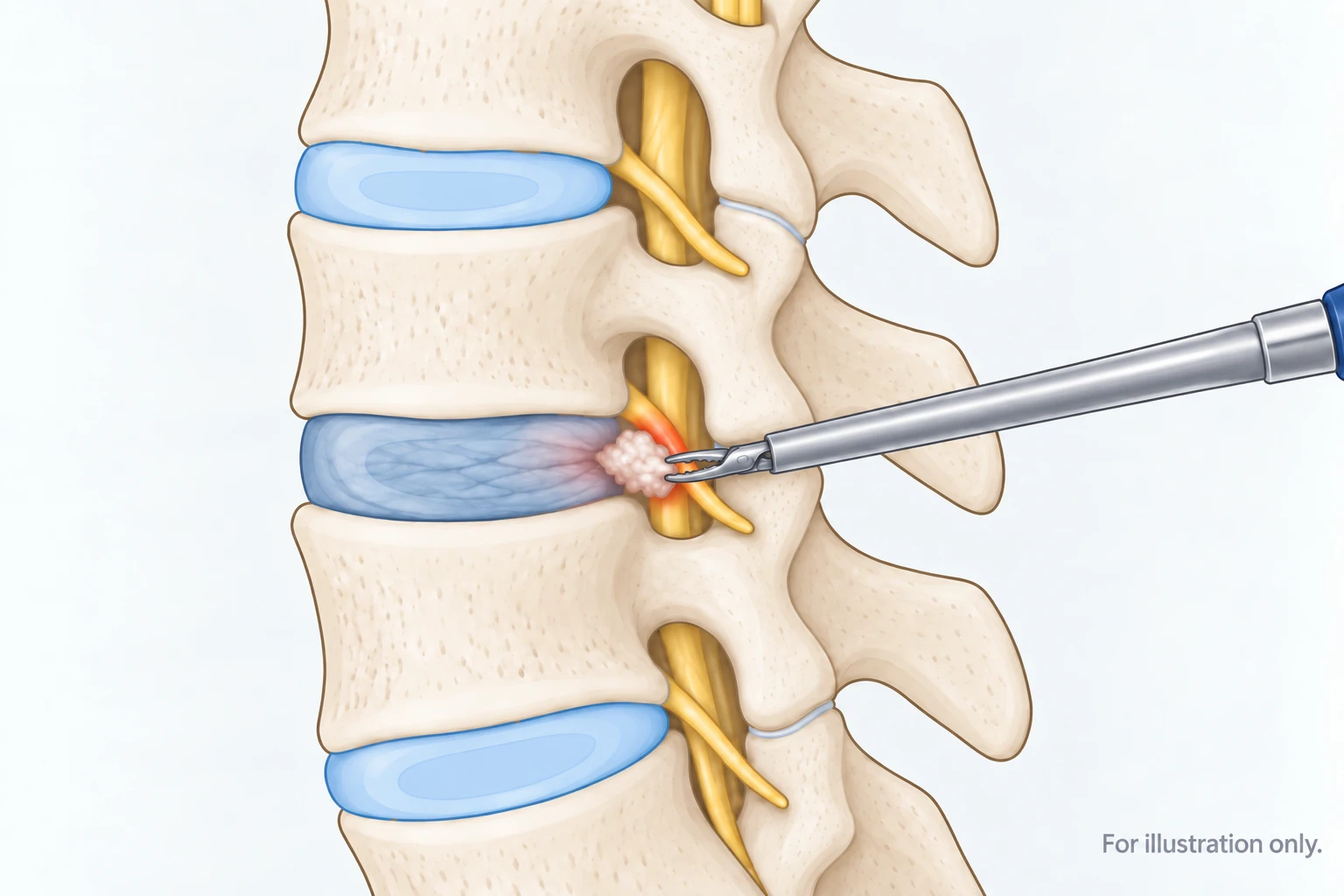
A microdiscectomy, laminectomy or laminotomy are common operations recommended for stenosis. The surgeon removes whatever is crowding the nerves, whether overgrown bone, thickened ligament, or a piece of bulging or herniated disc, which relieves the leg symptoms of claudication. For many people this is all that is needed. Recent studies that compared the two have found that adding a fusion does not reliably improve outcomes for stenosis without significant instability, or even for many people with a mild slip, so decompression on its own is frequently the right choice.

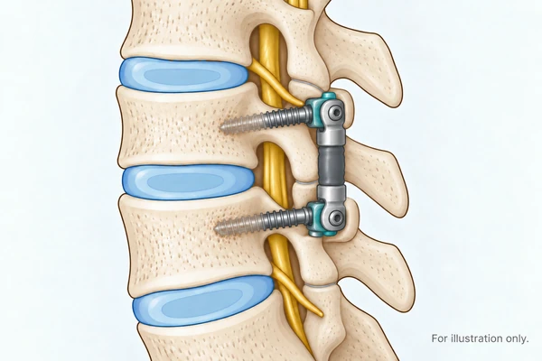
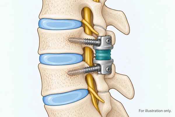
2. Dynamic stabilisation (HPS 2.0)

Where the segment genuinely is unstable and needs support, that support does not have to be a fusion. After the decompression, a dynamic stabilisation system can hold the level while still allowing it to move. Our German specialists use the HPS 2.0 system, which is designed for exactly this setting: degenerative stenosis, including a low-grade slip, following decompression. It preserves motion at the level rather than locking it.

3. Facet Joint Replacement (TOPS)

Freeing the nerves in stenosis sometimes means removing the facet joints themselves, and once they are gone the segment needs something to replace the role of these joints. The TOPS System is a facet joint replacement system built for this. After the decompression, it is anchored to the vertebrae with pedicle screws, and a central moving mechanism takes over the work of the facet joints, holding the level stable while still letting it bend, straighten, lean, and rotate. Where the dynamic stabilisation above supports the segment, TOPS fully replaces the joints that were removed.

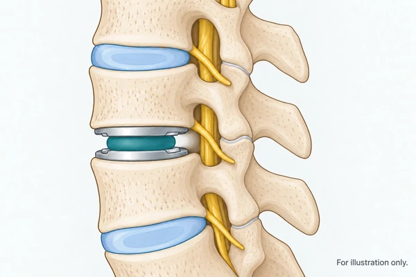
4. Artificial Disc Replacement (ADR)

When a degenerative disc loses height, the nerve tunnels called foramen can become narrowed and cause stenosis. Replacing the disc (and disc height) with an appropriately sized disc implant can open up these tunnels again and decompress the nerves.

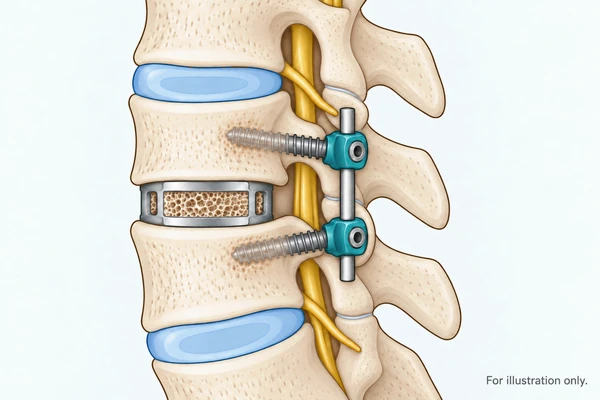
5. Spinal Fusion

Reserved for cases with significant instability where a motion-preserving option is not suitable. It stops movement at the level permanently, so it is not the first choice where something less will do the job.

In every case, the aim is to do enough to free the nerves and stabilise the spine where needed, without doing more than the problem calls for.

Nine years on from lumbar disc replacement in Germany (active and thriving)

Joel, from Auckland, New Zealand, had lived with back trouble for nearly thirty years, since an injury in his twenties. By early 2017 the wear had reached two levels of his lower back, L4/L5 and L5/S1, where disc degeneration and spinal stenosis, the narrowing that crowds the nerves, had developed together. When he sought advice at home, he was told he needed a double spinal fusion, and he wanted to avoid one if he could.

He came to us for a second opinion. After our surgeons reviewed his imaging, he was accepted for motion-preserving disc replacement at both levels, freeing the trapped nerves and replacing the worn discs rather than fusing the segments. He travelled to Germany with his family and had his surgery with Dr. Bierstedt in 2017. The day after surgery he was stood up with the back pain he had arrived with already gone, and he flew home a little over two weeks later.

Joel committed to his rehabilitation, working with a physiotherapist and an exercise physiologist to rebuild his strength and move well in daily life. Within months he was running and back in the gym, and within a year he completed a 23-kilometre alpine crossing. Nine years on, he is living an active life, family trips to Europe, regular training, and fishing trips through the Pacific, and we remain in regular contact.

What we see at Spine Connection

Many of the stenosis patients who reach us have already been told they need a spinal fusion. Often, once the imaging is reviewed, a decompression on its own, or a motion-preserving option where the segment genuinely needs support, is worth considering first. Our aim is to match the operation to what the spine actually needs.

Dr. Thomas Bierstedt is a neurosurgeon and spine surgeon at ONZ International in Germany, and a member of EuroSpine and AO Spine. He has spent much of his career on motion-preserving alternatives to spinal fusion, in both the lumbar and cervical spine.

Frequently Asked Questions

There is no single best one, and medication is a supporting measure rather than the treatment. Simple anti-inflammatories help some people with the inflammatory component. Neuropathic pain medications are sometimes used where leg symptoms are prominent. What works better than any tablet is the combination of staying active, physiotherapy including forward-bending exercises and walking programs, and weight management. Medication choice should be discussed with your own doctor.

Walking is worth persisting with, but the way you do it matters. Walking upright often brings on the symptoms, while leaning forward opens the space around the nerves and relieves them. This is why many people find they can walk much further leaning on a shopping trolley, or cycle comfortably when walking is difficult. Using those positions to keep walking is better than stopping.

It varies enormously and it is the measure most people use to track their own condition. Some walk for miles with occasional heaviness; others find symptoms come on within a few minutes. What is more useful than the distance itself is whether it is changing. A walking distance that is steadily shrinking, or leg weakness that is worsening, is the signal to have it reviewed.

The movements that tend to aggravate lumbar stenosis are the ones that arch the back backwards, such as standing extension, overhead lifting, and long periods standing still. Forward-bending positions usually relieve symptoms, which is why cycling and using an inclined treadmill are often better tolerated than walking upright. This is general guidance and a physiotherapist should tailor it to your case.

Age-related wear that narrows the space around the nerves in several ways at once. Discs dry out and lose height, bulging backwards and bringing the vertebrae closer together. Facet joints become arthritic and enlarge, crowding the canal. The ligamentum flavum thickens and buckles inward. Often several act together. Some people are simply born with a narrower canal, which means less age-related change is needed before symptoms appear.

For most people it is limiting rather than dangerous, and symptoms often stay stable or fluctuate rather than steadily worsening. It is not life-threatening. The situations that need urgent care are loss of bladder or bowel control, numbness around the groin or inner thighs, or leg weakness that is quickly worsening. These can signal cauda equina syndrome, which should be evaluated by a Spine Specialists as soon as possible.

No. Most people are managed without it, at least to begin with, and clinical guidelines recommend starting with conservative care. Surgery is considered when leg symptoms and walking limitation persist despite that, or where there is significant or worsening weakness.

Been told you need a spinal fusion?

If you have been told you need a fusion for spinal stenosis, or you are stuck on a waiting list, or facing a cost you cannot meet at home, it is worth getting a second opinion before you commit. Our specialists in Germany and Thailand can review your imaging and tell you honestly whether a decompression on its own, or a motion-preserving option, fits your case, or whether a fusion really is the right choice for you.

Sources

  1. Spinal Stenosis and Neurogenic Claudication. StatPearls (updated 2023). (Narrowing of the central canal, lateral recess, or neural foramen; part of the ageing process; neurogenic claudication.) (https://www.ncbi.nlm.nih.gov/books/NBK430872/)
  2. Katz JN, et al. Diagnosis and Management of Lumbar Spinal Stenosis: A Review. JAMA, 2022. (Neurogenic claudication, diagnosis, and management, including surgery for those not improving with conservative care.) (https://pubmed.ncbi.nlm.nih.gov/35503342/)
  3. Non-Surgical Interventions for Lumbar Spinal Stenosis Leading to Neurogenic Claudication: A Clinical Practice Guideline. The Journal of Pain, 2021. (https://www.jpain.org/article/S1526-5900(21)00188-7/fulltext)
  4. Kaiser R, et al. Decompression alone versus decompression with instrumented fusion in the treatment of lumbar degenerative spondylolisthesis: a systematic review and meta-analysis of randomised trials. J Neurol Neurosurg Psychiatry, 2023. (Adding fusion to decompression did not improve outcomes for degenerative spondylolisthesis.) (https://www.ncbi.nlm.nih.gov/pmc/articles/PMC10359551/)
  5. Lumbar dynamic pedicle-based stabilization versus fusion in degenerative disease: a multicentre, double-blind, randomized controlled trial. J Neurosurg Spine, 2022. (Category-level reference for the dynamic-stabilisation approach; not a study of the HPS 2.0 device.) (https://thejns.org/spine/view/journals/j-neurosurg-spine/37/4/article-p515.xml)
We're here
to help.
Leave your details and we will contact you to answer your questions.

Get a Spine Specialist's Opinion for Your Spinal Stenosis

Leave your details and we will contact you to answer your questions. We're here to help.


    Join Our Youtube Channel

    Watch videos showcasing the latest technologies and surgery techniques, and keep up to date with patient stories from around the globe.